Getting a breast cancer diagnosis involves many imaging tests, but Positron Emission tomography (PET) Scans are not part of the standard of care for diagnosing early or locally advanced breast cancer since they may miss small tumors. There are myths when it comes to PET scans for breast cancer, but PET scans can be helpful when standard imaging results are unclear or when looking for signs of metastasis (cancer spread). PET is also being studied for its role in staging breast cancer and detecting cancer in underarm lymph nodes (axillary lymph nodes). PET scans are most commonly used for metastatic breast cancer (MBC) to assess disease extent, evaluate treatment response and help guide personalized care.

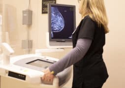
PET scans are a type of molecular imaging test, meaning that they provide information about how the body functions rather than its structure, like X-rays, CT scans and MRIs provide.
During a PET scan, a radioactive molecule called a radiotracer (tracer) is given through a vein (IV). The tracer travels through the blood looking for anything abnormal. A PET scanner takes images of the body where the tracer has been absorbed by cancer cells which have collected in tissues and organs. PET scans are usually combined with CT scans. When the images are combined, a radiologist has a good idea about where the cancer is and how it’s behaving, even if multiple tumors are present.
It is important to understand the powerful role that PET scans can have in your breast cancer experience. There are four common myths about using PET scans:
Myth: All PET scans are the same.
There are two different types of PET scans used for breast cancer that address different needs:
- FDG-PET scans measure the metabolic activity of a tumor, as high metabolic activity can translate into a tumor that is more aggressive. This means this type of scan can be used to look for MBC and determine if a treatment is working and also to detect recurrence in some cases.
- FES-PET scans may be an option for some patients with MBC, ER+, or recurrent breast cancer to determine if the metastatic tumors are positive for the estrogen receptor. This can help doctors plan the best treatment.
Myth: Everyone needs a PET scan.
PET scans are helpful in some cases but are not necessary for everyone. Since FDG-PET scans are primarily used to assess how tumors have grown and spread, they are not typically needed for those with early-stage breast cancer. However, for those newly diagnosed with stage 3 breast cancer, this type of PET scan may be an option at the time of diagnosis, depending on symptoms and other test results.
For those with MBC or locally recurrent breast cancer, both FDG-PET and FES-PET scans may be used. FDG-PET evaluates the spread of cancer cells, while FES-PET determines if metastatic tumors are estrogen receptor-positive, which can help guide treatment decisions.
Myth: PET scans expose you to too much radiation.
Everyone is exposed to small amounts of radiation daily from natural sources. While PET scans do involve radiation, the exposure is carefully controlled and considered safe. FES-PET scans have slightly lower radiation exposure than FDG-PET/CT scans, which include a CT component that adds to the total dose. For context, the radiation from a PET scan is roughly equal to several years of natural background exposure.
When deciding whether a PET scan is right for you, factors like the type of scan, the information it provides and how it will guide your treatment should be considered. If you have concerns about radiation risks, talk to your doctor — they can help you weigh the benefits of a PET scan in your specific situation.
Myth: PET scans can replace all other imaging tests.
PET scans provide valuable information about how cancer behaves, but they do not replace other imaging tests.
Mammograms detect abnormalities in the breast. If your screening mammogram shows something abnormal, you’ll need follow-up tests, like a breast ultrasound. Breast MRIs are used to screen women at high risk of breast cancer, as well as in diagnosis and staging. They provide highly detailed images of breast tissue. CT scans help determine if cancer has spread to organs such as the lungs or liver. Bone scans are used to detect cancer that has spread to the bones.
PET scans are most useful for staging metastatic breast cancer and assessing treatment response. They work best when used alongside other imaging tests, not as a replacement. If you have questions about which imaging test is right for you, talk to your doctor about how different scans can provide the information needed to guide your care.
If you’re facing a breast cancer diagnosis and not sure where to begin, you’ve come to the right place. Our Know More series will continue to help educate you and arm you with the tools you’ll need to feel empowered to advocate for yourself throughout your experience.
Read More
Downloadable Resource: Types of Imaging Scans and What They Show
Tests for Metastases in People Newly Diagnosed with Breast Cancer
Hear More:
Real Pink Podcast: A Gateway to Empowerment: What to Know About Molecular Imaging
Imaging and Future of MBC Treatment with David Mankoff
More Actions You Can Take:
The Komen Patient Care Center is your trusted, go-to source for timely, accurate breast health and breast cancer information, services and resources. Our navigators offer free, personalized support for you and your loved ones including education, emotional support, financial assistance, help accessing care and more.
Join ShareForCures®. ShareForCures® is Komen’s breast bancer research registry that will answer some of the most pressing questions in breast cancer. Anyone diagnosed with breast cancer over age 18 in the U.S. is eligible to participate.
Take this survey to let us know what you think of this content as part of the Know More Series.
This blog is sponsored by: