Mammograms and more may be part of your breast cancer screening plan, depending on your personal risk. Finding breast cancer early makes a big difference for treatment options and long-term survival.
Breast cancer screening tests are designed to find breast cancer before warning signs or symptoms appear, and different types exist to clarify uncertain findings – offering peace of mind or helping inform next steps. For most women at average risk, screening begins with a yearly mammogram at age 40. The screening tools recommended for you depend on factors such as your age, family history, personal history and breast density. Mammograms remain the most common breast cancer screening test, but they are not the only imaging tool your doctor may recommend. You might also hear about breast MRI, breast ultrasound or newer imaging tests under study. Here’s a look at the most common options and what makes them different – starting with a mammogram.
Clinical breast exams and mammography: the foundation of breast cancer screening
A clinical breast exam (CBE) and mammography form the foundation of routine annual screening for people at average risk. CBE is a physical exam in which a health care provider looks at and feels your breasts and underarm areas for anything unusual. Mammography uses low-dose X‑rays to create images of the breast, called mammograms, which can find signs of cancer before it can be felt.
You may hear about 2D or 3D mammography. Traditional 2D mammography takes a flat image of the breast from two different perspectives or angles (from the front and the side). It’s been used for decades and helps flag abnormalities in the breast, but it has limitations – especially for people with dense breast tissue, where cancers can be harder to spot.
3D mammography, also known as digital breast tomosynthesis, takes many X-ray images from different angles. It constructs a stack of these thin, layered images – like flipping pages through a book. This way, a doctor can examine breast tissue layer by layer, opposed to a flat image, which has helped to find more cancers than 2D mammography, particularly in dense breast tissue. It also reduces the number of callbacks for additional imaging. This method is now the standard approach used by most mammography centers in the U.S., so if you recently had a mammogram, it was likely a 3D screening.
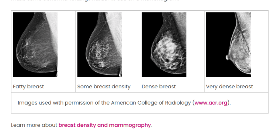
Why breast density matters for screening
Breast density refers to how much fibrous and glandular tissue is in the breast compared to fatty tissue. This is determined by a mammogram, not by how your breasts may look or feel. About half of women who get mammograms have what is considered dense breast tissue. Breast density matters for two reasons. One already mentioned is that it can make it harder to read mammograms because dense tissue and tumors both appear white on the image, which can be like trying to find a polar bear in a snowstorm. The other reason is that having dense breast tissue increases your risk of breast cancer.
Breast density is one reason your doctor may suggest additional screening because it can help reveal more information about your breast health. Additional screening could include some of the next few tools discussed.
Breast magnetic resonance imaging (MRI) is a useful tool for some higher-risk women
MRI uses magnets to create images of the breast structure. Breast MRI is not part of routine screening for most people, but it’s recommended alongside mammography for some at higher risk, like those with certain inherited mutations (BRCA1/2), prior chest radiation earlier in life or because of family history of breast or other cancers. Also, mammography plus breast MRI is under study for screening in women with dense breast tissue. In people with dense breasts, breast MRI plus mammography may find a few more breast cancers than mammography alone.
Breast MRI can result in more false positives, meaning that it may detect something benign or suggest something is there that actually is not. It’s also not available at every care center, and some people may need to travel to access it. However, it can provide supplemental information to help your doctor understand your situation. Some people who need long-term screening may alternate between mammograms and breast MRIs annually, so they don’t have to get an MRI as frequently. But first, talk with your doctor. Together, you can make a screening plan that’s right for you.
Breast ultrasound gives a closer look
Breast ultrasound uses sound waves to make images of the breast. It is not a replacement for mammography but instead used as a follow-up test after an abnormal mammogram, MRI or clinical breast exam. This is called targeted ultrasound because it focuses on one specific area of concern.
In some cases, whole-breast ultrasound may be used to look at more tissue, especially for those with dense breast tissue or those who cannot have a breast MRI. Breast ultrasound is not used as a screening tool alone or to replace mammography and can lead to more false positives. However, mammography combined with whole breast ultrasound is under study to see if they are better combined than mammography alone in finding breast cancer, as it helps provide another visual perspective of the tissue.
Other emerging screening tools may be used in certain situations
Women at higher risk of breast cancer who are recommended to have breast MRI as part of their breast cancer screening, but cannot have one for medical reasons, may consider contrast-enhanced mammography (CEM) or molecular breast imaging (MBI) – and if these are not available, whole breast ultrasound.
MBI and CEM are newer tools under study. MBI uses a small amount of radioactive tracer, and special cameras help highlight where cells are more active. The goal of this tool is to find cancers that are harder to see on a mammogram as it shows how much cells are metabolizing, and active cancer cells will take up more tracer with a higher rate of metabolism.
CEM is a type of mammogram that uses a small injection of contrast dye. It shows a stronger visual contrast between a breast tumor and surrounding tissue than your typical mammogram. CEM is another option that’s particularly helpful in some cases like when screening dense breast tissue, and it can help provide information similar to contrast-based imaging though it is not the same as a breast MRI.
Your doctor will help you understand your risk and the screening plan that fits you best
Breast cancer screening is not one-size-fits-all, and depending on factors like risk and breast density, a doctor might recommend different screening options. These tools can provide more information and more peace of mind that comes with that. Learn more about how to understand your risk and how to discuss it with your doctor so together you can create a screening plan that works best for you.
Content covered in the Know More Educational Series may be an emerging area in research or technology. Talk with your doctor about what is right for you.
We’d love your thoughts!
Take our brief survey and browse the rest of the Know More series here.
Read More:
Know Your Risk: Educate, Empower, Act blog
What You Need to Know About Imaging for Breast Cancer Screening
Mammography recommendations for:
Watch More:
The Importance of Knowing if You Are High Risk
MBC Impact Series – The Importance of Imaging in MBC Treatment
What Happens if You Get a Call Back After a Mammogram?