Nuclear medicine is a specialized field of imaging that uses tiny amounts of radioactive substances to detect and monitor diseases, which can help shape breast cancer care. Unlike traditional imaging methods like X-rays, MRIs or ultrasounds, which create pictures of the body’s structures, nuclear medicine shows how cells and tissues function at a molecular level. Below are five ways this deeper look can help doctors spot breast cancer earlier, see if it has spread and track how well it is responding to treatment.
How nuclear medicine plays a role in breast cancer
At different stages of a breast cancer journey, nuclear medicine can provide valuable insights that help shape care decisions. PET scans, bone scans and lymph node scans do more than just observe the cancer — they help doctors determine the best course of action, whether planning surgery, selecting the right medications or monitoring treatment effectiveness.
To truly appreciate the role of nuclear medicine, it helps to see it in action. The following real-world scenarios were provided by the Society for Nuclear Medicine and Molecular Imaging’s Breast Imaging Outreach Working Group and illustrate how these imaging techniques provide critical information that guides diagnosis, staging and treatment decisions and how nuclear medicine may be used in different breast cancer situations. Every person’s experience is unique, and care decisions should always be made with your health care team.
Bone scans
Jane is a highly active woman with a new diagnosis of a larger low-grade estrogen receptor-positive (ER-positive) cancer with three positive axillary lymph nodes. She has noticed a recent onset of mid-back pain unrelated to an injury or exercise. Her doctor orders a bone scan to help determine if the cancer has spread (metastasized) to her bones and to investigate the cause of her pain. The scan reveals an area of abnormal activity in her mid-back. She then undergoes an MRI of her spine followed by a breast biopsy, which confirms ER-positive metastatic breast cancer. With this information, Jane’s doctor changes her treatment plan to include systemic therapies, such as hormone therapy, to target the breast cancer throughout her body together with bone-targeted medication instead of focusing solely on local treatment.
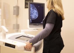
Molecular Breast Imaging (MBI)
Mary is 40 years old and recently had a screening mammogram. The mammogram showed a suspicious finding in her left breast. The report also noted that she has dense breast tissue, which may make mammograms less effective at detecting cancer in very dense breast tissue. Since Mary is unable to tolerate an MRI, her doctor recommends molecular breast imaging. Molecular breast imaging (MBI) is a specialized test that may provide a clearer view of some cases of dense breast tissue. The scan revealed an additional suspicious area in her left breast that was not visible on the mammogram. A follow-up breast biopsy confirms this area is an additional site of cancer. With this information, her doctor recommends the most appropriate surgical option for Mary, opting for a mastectomy instead of a lumpectomy to ensure all the cancerous tissue is removed.
FES-PET/CT
Anna was diagnosed with estrogen receptor-positive breast cancer 15 years ago. When she developed persistent bone pain, her doctor ordered a bone scan, confirming her cancer had spread. To guide treatment, her doctor performed an FES-PET/CT scan, which is an option for some people with metastatic or recurrent estrogen receptor-positive breast cancer as it can detect estrogen receptors in cancer cells. The results showed her metastatic tumors were no longer ER-positive, meaning hormone therapy would no longer be effective. This crucial information allowed her doctor to switch to more suitable treatments, such as chemotherapy or targeted therapy, and consider a biopsy for further guidance.
Serial FDG-PET/CT
Luisa has metastatic triple negative breast cancer. To determine where the cancer had spread, her doctor used an FDG-PET/CT scan, which highlights cancerous areas by detecting a special radiotracer (FDG) absorbed by cancer cells. After starting chemotherapy, Luisa had a follow-up FDG-PET/CT scan, which showed that many of her tumors had shrunk and absorbed less FDG, indicating that the treatment was working. This gave Luisa and her doctors the confidence to continue with her current chemotherapy plan.
Sentinel node mapping
Christina was diagnosed with a small invasive breast cancer found during a routine mammogram. Her doctor found no signs that the cancer had spread to her lymph nodes through a physical exam or breast ultrasound. To be sure, her surgeon performed a sentinel lymph node biopsy, a procedure that identifies the first lymph node(s) where cancer would likely spread. A small amount of radioactive tracer was used to locate the nodes, which were then removed and tested. The results showed no cancer in the sentinel nodes, meaning Christina avoided more extensive lymph node surgery. This reduced her risk of complications, including lymphedema, a condition that can cause arm swelling.
Talk to your doctor to learn which imaging tools or tests may be right for your situation. Your care team can help determine what’s best for you based on your diagnosis and treatment goals.
Timeline of when nuclear medicine is used in a breast cancer
Understanding when nuclear medicine may come into play can help you feel more informed and prepared. It may be used:
- At diagnosis – Can be used to evaluate some cases of dense breast tissue, confirm suspicious findings or assess if the cancer has spread.
- For staging – To determine whether cancer has spread to the lymph nodes or bones.
- To guide treatment – To check for hormone receptor status or monitor how well treatments are working.
- For detecting recurrence – To identify cancer returning earlier than standard imaging methods can.
To help visualize where these imaging techniques fit into a breast cancer timeline, download our Nuclear Medicine and Breast Cancer Guide Downloadable Resource.
Content covered in the Know More Educational Series may be an emerging area in research or technology. Talk to your doctor about what is right for you.
We’d love your thoughts!
Take our brief survey and browse the rest of the Know More series here.
Learn More:
Molecular Breast Imaging Fact Sheet
Precision Imaging for Breast Cancer